Matron’s Medical Report Book – Part 1

The Matron’s Medical Report Book is the earliest surviving record we have from Stannington Sanatorium, which contains monthly updates on patient progress and general administration.  The first entry from the Report Book, below, dates from 1908 and lists some of the earliest patients with details of their ages, addresses and condition whilst in the Sanatorium.    


April 11th 1908

“There are now six patients here.  Five have been here for nearly four weeks (since March 18th) & one for three weeks.  They are:-

1. Maggie Smith, aged 17.  Address 73 Leopold St (sent from the Gateshead Workhouse)

2. John Edward Kenney, aged 15 ¾; Address 72 William St, Hebburn Quay (working at Hebburn Colliery)

3. James Robson, aged 13. Address 16 Bottle Bank, South Shields

4. Thomas Hill, aged 12 ½. Address 76 Belvedere St, Heaton

5. John Nicholson, aged 10.  Address 10 Hyiamais Court, Gateshead

6. Thomas Burns, aged 16 ¾. Admitted March 21st. Address 40 Stanley St, Jarrow


The general condition of all the patients has considerably improved.  They have all gained in weight during the last 18 days: Maggie Smith 5 ¼ lb, John Kennedy 4lb, James Robson 2 ¾ lb, T. Hill 3lb, J. Nicholson 3 ¾ lb, T. Burns 3 ¼ lb (all given to the nearest ¼ lb).  Only one patient, Maggie Smith, has any marked degree of fever.  There is a slight improvement in her temperature.  One patient, J. Hill, does not cough at all.  All the others are coughing less and bringing up less sputum.

Examination of the sputum for tubercle bacilli has not yet been possible.

The local condition in three patients, T. Burns, T. Hill & J. Nicholson is quite satisfactory, shewing improvement. In two patients, Maggie Smith & J. Robson it has remained about the same.

In one, J. Kennedy, there has been some extension of the diseased area, but improvement again during the last four days after strict rest.”

[HOSP/STAN/2/1/1]


The close association of tuberculosis with poor living conditions is further highlighted here by the first two patients who have come from Gateshead Workhouse and Hebburn Colliery respectively.  At this stage there were only 6 patients but the sanatorium had opened not long before this with provision for 40 children and soon saw the beds fill up.  In the following years the sanatorium’s capacity began to expand rapidly with an extension providing an additional 50 beds opening in 1911 and several new wards opening in the 1920s so that by 1926 there were 312 beds available.

HOSP-STAN-9-1-1 aerial view

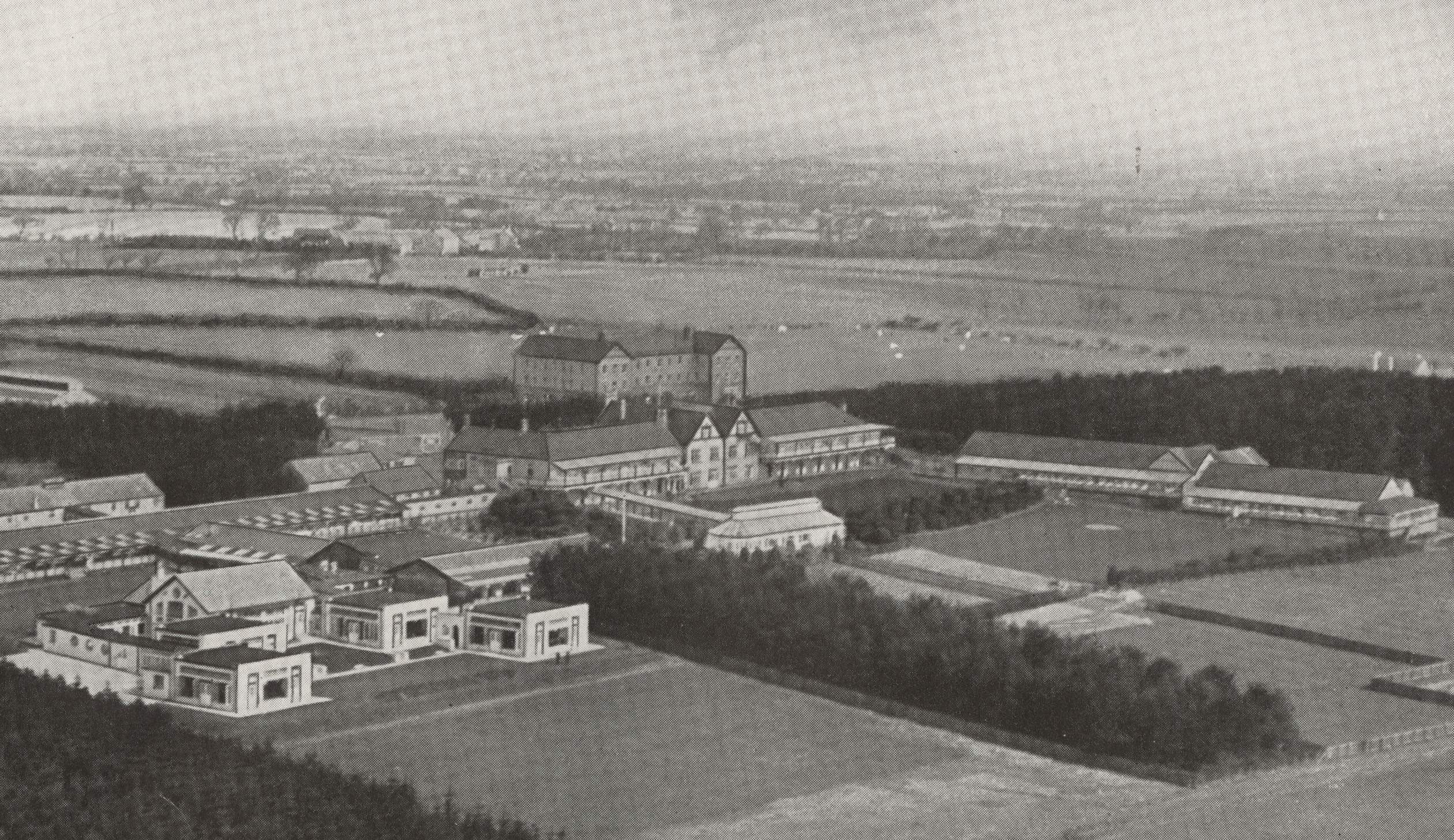
Aerial view of the sanatorium c.1936

[HOSP/STAN/9/1/1]

We will post some more entries from the Matron’s Report Book in future blog posts to track the progress of the early days of the Sanatorium.

Patient Files

As part of the Stannington collection we have patient case files spanning the years 1939-1966 containing a wealth of medical and social information to support that found in the radiographs.  The earlier files have a different format to the later ones owing to a change in the administration of patient records at Stannington which occurred in 1946.  Up to 1946 the patient records take a much larger format and the patients were all allocated their own unique patient number based on their date of discharge, whereas from 1946 onwards standard sized paper files come into use with patient numbers being based on date of admission.

[HOSP/STAN/7/1/1/1587]

The above file is an example of one of the later files with the patient’s name redacted for confidentiality purposes.  Three different colour files were used, each one indicating the type of tuberculosis the patient was suffering from.  Blue files were used for sufferers of pulmonary TB, pink files for non-pulmonary TB, and finally green files for TB of the bones and joints.  This image gives a good indication of the sort of information that can be found on the files, which is also indicative of the information we will be recording in our catalogue.  The information featured in the catalogue for each patient will be as follows: patient number, date of admission, date of discharge, sex, age on admission, home town, diagnosis, result of treatment, where admitted from, the local authority sending them, and where applicable any re-admission numbers and dates.


To clarify some of the information given on the file, the date of immunisation refers to immunisation against diphtheria, not tuberculosis, as widespread vaccination against TB was not yet in place.  As a contagious disease and a major concern for public health, all diagnosed cases of tuberculosis had to be made known to the local public health authorities, which is what the notification date refers to.


Each case of tuberculosis had to be classified according to centrally issued guidelines and this is often noted on the patient’s file under diagnosis. The first distinction made when classifying the disease is between pulmonary and non-pulmonary tuberculosis, with pulmonary including TB of the pleura and intrathoracic glands and any patient suffering from a combination of pulmonary and non-pulmonary TB would be classified as pulmonary.  Cases of pulmonary TB could then be subdivided between TB minus and TB plus.  Cases in which tubercle bacilli have never been found in the sputum or other pathology samples are classed as TB minus, as the above patient is.  TB plus on the other hand applies to cases in which tubercle bacilli have at some point been found and is subdivided further into 3 groups; group 1 applying to cases with slight constitutional disturbances if any and limited physical signs, group 3 for cases showing profound constitutional disturbance or deterioration and with little or no prospect of recovery, and finally group 2 for all cases which cannot be placed within groups 1 or 3.  Patients suffering from non-pulmonary tuberculosis would be classified according to the site of the lesion, for example, tuberculosis of the bones and joints, abdominal tuberculosis, tuberculosis of other organs, and tuberculosis of the peripheral glands.


There is also a space on each patient file to enter the result of treatment and there were also central guidelines covering this.  Most patients leaving Stannington are deemed to be ‘quiescent’, meaning that they have no signs or symptoms of tuberculous disease and any sputum is free of tubercle bacilli.  A patient’s condition could also be classed as ‘arrested’ by which it is meant that in pulmonary cases the disease has been quiescent for at least two years and in non-pulmonary cases it is quiescent and there is no reason to believe it will recur.  And finally, a patient could be considered to be ‘recovered’ if the disease had been arrested for at least three years.


The information that you can expect to find within the patient records does vary from patient to patient but generally includes data on other family members, living conditions, medical history, temperature charts, x-ray reports, pathology reports, details of progress, and any correspondence with family members or local authorities.  The correspondence contained within the files can give a fascinating insight into social problems and the impact tuberculosis could have on families at the time adding an extra dimension to the medical information that we expect to find.  The image below is an example of the x-ray reports that can be found in the back of some of the files; it is quite common for files dating from the mid-1940s to find small diagrams of what was seen in the x-rays also.


x-ray reports

[HOSP/STAN/7/1/1/1587]


Case Study – Pulmonary Tuberculosis

In this post we’re going to explore the progression of pulmonary tuberculosis in one particular patient from Stannington Sanatorium in order to gain an insight into some of the common approaches to the treatment of the disease at this time.


Patient 95/1947 was admitted to Stannington Sanatorium on 4th September 1947 at the age of 12. After having begun to feel ill earlier in the year she was examined at the local clinic and sent for x-ray whereupon it was determined that she should be admitted to the sanatorium for treatment.  Prior to admission she had been living with her mother, step-father, two younger brothers and one younger sister in a 3 roomed house in Cockermouth which had no inside water or inside toilet.  The only family history of TB had been her father who had died from the disease when she was still a baby.  On admission she had no cough but a very poor appetite and was losing weight, weighing only 4st 0lbs 6oz.  There were no other physical symptoms or abnormalities reported.


The report on her first x-ray taken 4 days after admission reads:

Tuberculous infiltration of both upper lobes with a large cavity in the mid-zone & a smaller one at the left apex.  There are several small calcified foci in the right upper lobe.”

Continuing reports over the next 4 months describe great improvement on the right side with the cavity in the right mid-zone no longer being visible.  However, the condition of the left side continues to deteriorate with a report 7 months after admission stating that the “cavity in the left upper lobe is now very much larger 1 ½” in diameter.”


NRO-3000-HOSP-STAN-07-01-02-1444-19

[HOSP/STAN/7/1/2/1444/19 – tomograph showing large cavity in left upper lobe, Dec 1948]


During her stay a series of different treatments were attempted to reduce the cavities.  Two months after admission in November 1947 her doctor initially observed that it was “doubtful if a satisfactory collapse could be obtained.  No treatment recommended.  Outlook very poor.”  Nevertheless, two months later in January an artificial pneumothorax was attempted but without success.


Artificial pneumothoraxes were performed on patients with the intention of resting the affected lung and hopefully collapsing the cavities at the same time whilst preventing any further spread as a collapsed lung was less likely to spread bacilli.  The procedure had been shown to effect a marked improvement in the size of tuberculous cavities for some patients but could at the same time be a dangerous procedure with a risk of air embolisms, pleural shock, sepsis, emphysema and effusion.


HOSP_STAN_9_1_1

 [HOSP/STAN/9/1/1, artificial pneumothorax treatment being performed in Stannington]


Three months later, after observing the growth of the cavity in the left upper lobe, a phrenic crush followed by a pneumoperitoneum was recommended and she was transferred to Shotley Bridge Hospital soon after for the procedures to be performed.  By crushing the left phrenic nerve, situated in the neck, they would be able to disable the left diaphragm thus forcing the muscle to relax and lift up, with the idea being that this would then rest the lower part of the lung.   A pneumoperitoneum was often performed in conjunction with the phrenic crush and involved inserting air into the abdominal peritoneal cavity forcing the diaphragm up.


Unfortunately after the patient was transferred to Shotley Bridge Hospital for the above procedures she never returned to Stannington and so we do not have any later case notes to follow up the result of her treatment.  However, some later correspondence does tell us that she was moved to Poole Sanatorium from where she was eventually discharged in May 1950.


The surgical procedures described here sound very drastic from a modern perspective but were a common approach in the pre-antibiotic era.  With no effective drug treatments surgical approaches such as these were at the forefront of tuberculosis treatment and looking through the files of Stannington Sanatorium it is clear that many of their young patients recovered, or at least showed significant improvements, and went on to live normal lives.