Tuberculosis of the bones and joints affected several key areas of the body, and is well documented amongst the Stannington records. Of these the knee is one of the more frequently noted areas of infection. Immobilisation by plaster cast was the most common form of treatment for this type of tuberculosis, although some more severe cases were put forward for surgical intervention.
Tuberculous arthritis characteristically affects only one joint, predominantly a weight-bearing joint such as the spine, hip or knee. It is transferred by haematogenous spread from a location of primary infection, most commonly the lungs. Initial symptoms often include synovitis or inflammation of the soft tissue in addition to joint effusion, where there is an increase in the fluid within the joint. These preliminary symptoms progress into arthritis over a period of time, although radiographic findings only begin to occur after three or four weeks. Ultimately, untreated tuberculous arthritis will lead to demineralisation, erosion and joint destruction.
Case Study

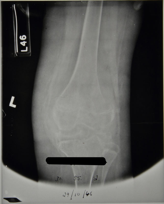
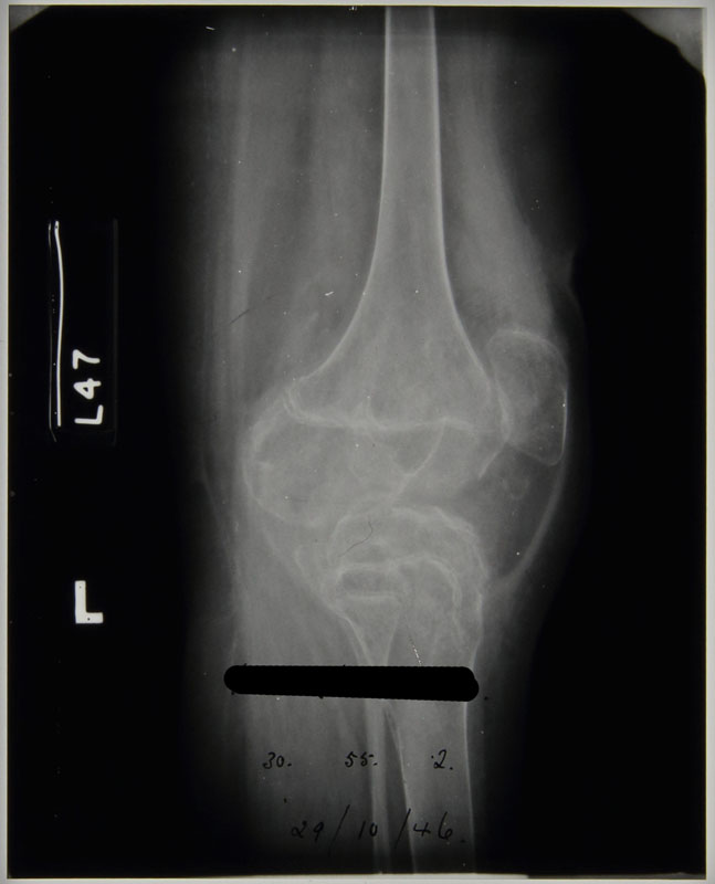
Patient 358/1946 was admitted to Stannington Sanatorium in October 1946 with tuberculous arthritis of the left knee. The patient notes detail that on admittance there was radiographic evidence of destructive lesions already identifiable, however, the first radiographs taken of the individual are of poor exposure or whilst the individual was in plaster cast, so identification is challenging.
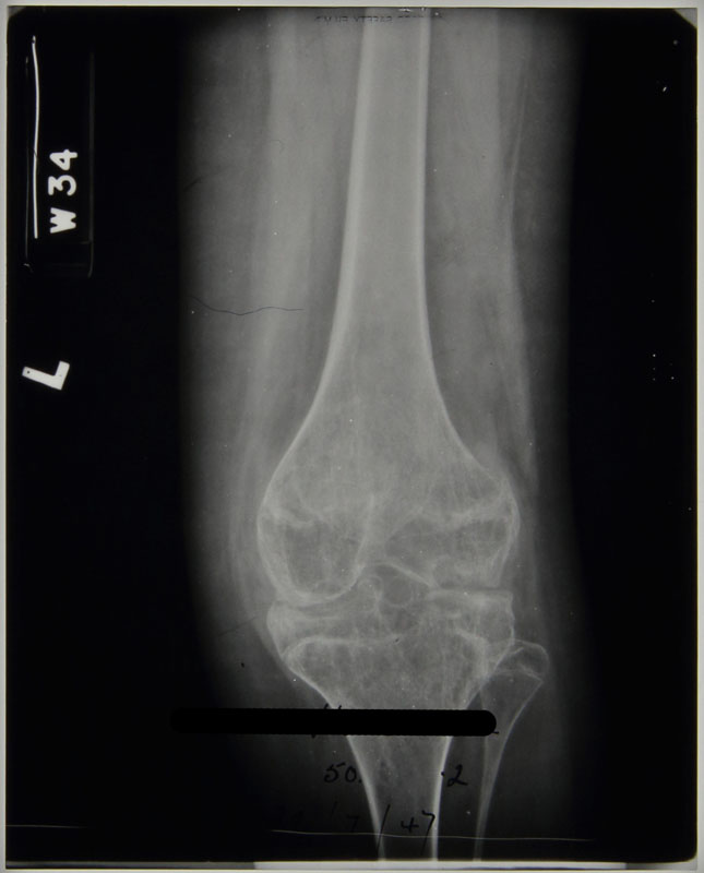
The radiographs from February 1947 show the bony anomalies to the knee joint clearly. There is a significant reduction in joint space between the femur and the tibia. The distal epiphysis of the femur shows severe displacement, having moved towards the posterior. Similar displacement can be seen on the proximal tibia to a slightly lesser degree. The patient notes at this stage indicate no change from time of admittance that two sinuses were present above the patella and that immobilisation of the knee was to continue.
In August 1947, an examination by the visiting physician describes:
‘Complete disorganisation of the joint. Less decalcification and bony trabeculae are beginning to show.
Fusion of the joint is not complete and there is still some heat.
To be put in plaster for three months’
Changes in the radiographic images between February 1947 and June 1948, when the patient is discharged, are minimal. In December 1947 the physician stated in the patient’s notes:
‘No change in appearance.
There is not complete bony ankyloses of the knee but movement is negligible.
A sinus on the front of the knee which is covered by a scab, is not at present discharging’
There is little or no heat in the knee.
For Thomas’ walking knee splint, patton and crutches.’
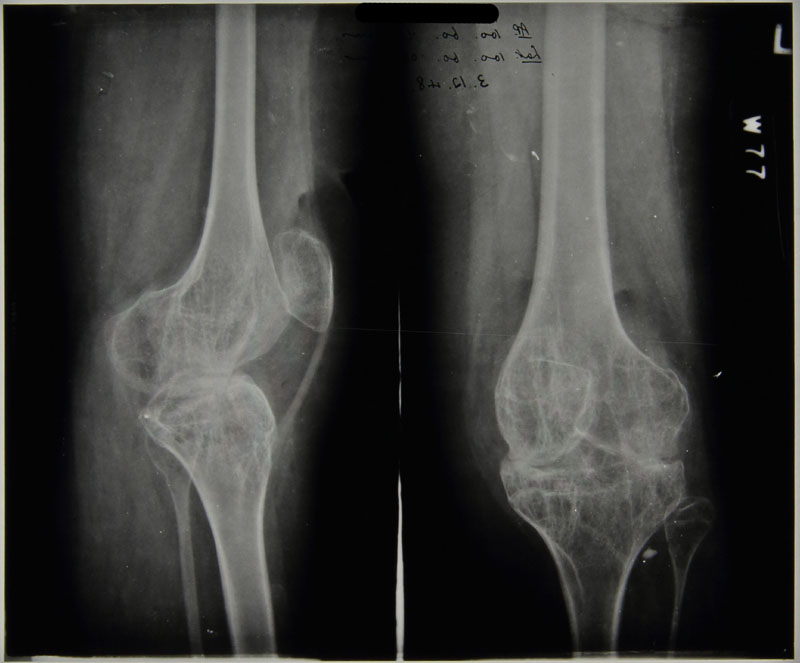
No further changes were noted at this stage with the radiographic image below, dated to December 1947, revealing gross anatomical destruction of the knee joint to have taken place and there is no remaining joint space. The striation pattern across the epiphysis and metaphysis of both the femur and tibia is likely to be the result of cartilage destruction and bone degeneration causing porosity in the bones.
Patient 358/1946 was discharged in June 1948 but according to their patient notes returned twice as an out-patient and was seen a further two times at the Sanderson Orthopaedic Hospital, Gosforth.
For a case study on the surgical interventions used in tuberculosis of the hip, see earlier post of 08/12/2014
Further radiographic images can be seen on Flickr at https://www.flickr.com/photos/99322319@N07/sets/72157648833066476/
Sources
Albuquerque-Jonathan, G (2006). Atypical tuberculosis of the knee joint. South African Journal of Radiology p.28.
Arthanari, S; Yusuf, S and Nisar, M (2008). Tuberculosis of the Knee Complicating Seronegative Arthritis. Journal of Rheumatology: http://www.jrheum.com/subscribers/08/06/1227.html