A number of the patients admitted to Stannington were mistakenly given an initial diagnosis of tuberculosis or were found upon examination to be non-tuberculous and were instead allocated an alternative diagnosis. Perthes’ Disease was the most common differential diagnosis assigned to the bones and joints in Stannington, affecting the hip joint this condition was often mistaken as tuberculous-arthritis of the hip.
Perthes’ Disease, a condition that usually affects children between the ages of 4 and 10, is a condition where the blood supply to the femoral head is temporarily lost. This causes the bone at the epiphysis of the femur to soften and breakdown, known as necrosis, giving the femoral head a flattened appearance.
Patient 88/38, a 6 year old boy, is one example of a patient from Stannington with Perthes’ Disease, in this case affecting the left hip. Admitted to Stannington on 31st October 1941, clinical notes read:
‘L. hip – some wasting thigh muscles. Some limitation flexion. Hip in good position.’

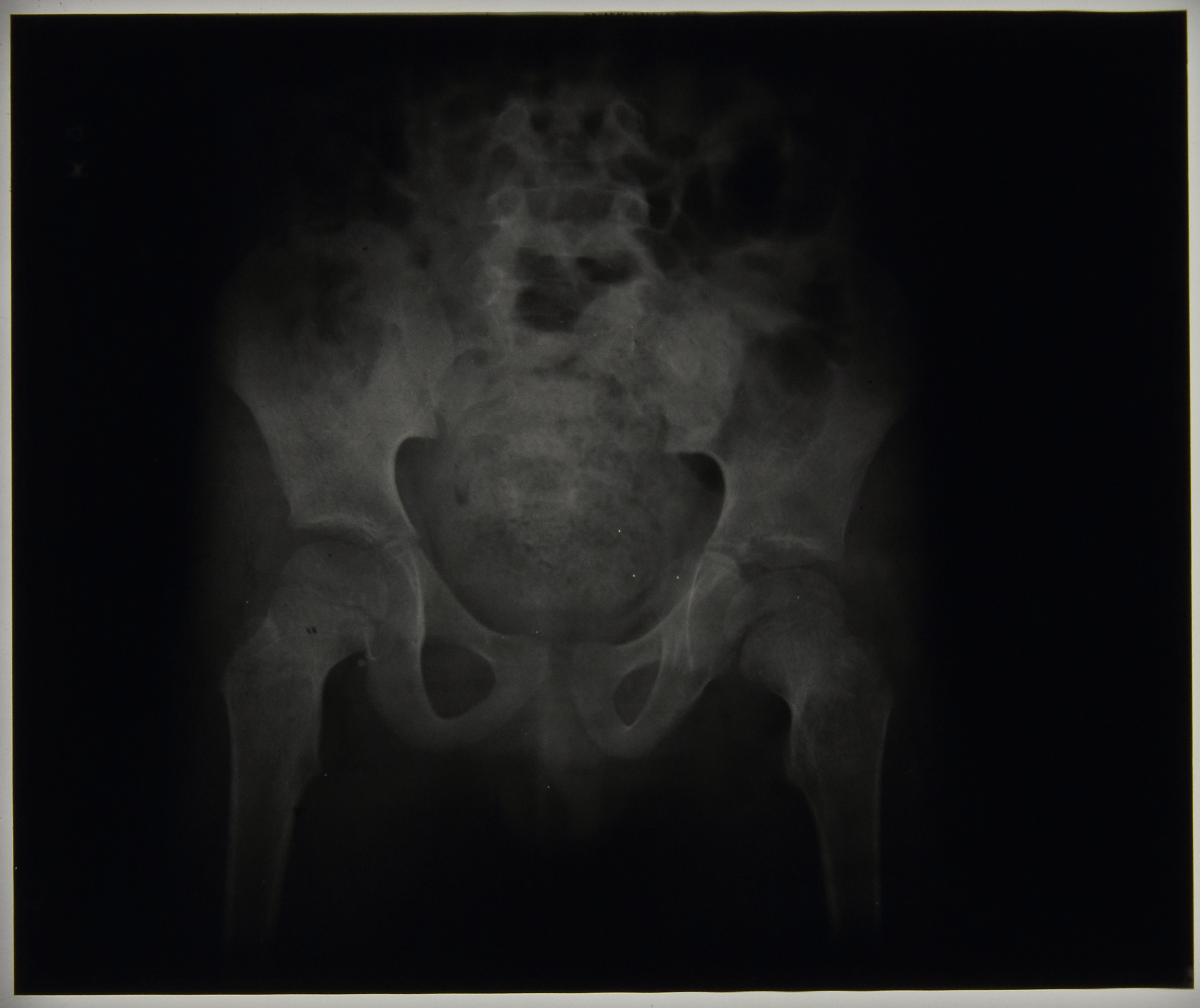
This is supported by the radiographs for the patient, figures 1 and 2 respective of date, and x-ray report:
‘13/11/1941 – Hip – flattened epiphysis has progressed since last x-ray 15/09/1941. Perthes’ Disease
20/11/1941 -Marked flattening of epiphysis L. hip. Some softening of neck. Definite Perthes’ Disease.’
By comparison, tuberculosis of the hip (see posting from 08/12/2014) results in the gradual destruction of the hip joint beginning with a reduction in the joint space between the femoral head and the acetabulum, leading to possible porosity and eburnation in the affected bones and the possibility of pathological dislocation, deformity and loss of use in the affected joint. Even after the disease has reached quiescence it is possible that the individual will suffer with ongoing osteoarthritis or ankyloses.

Figure 2, above, is of patient 88/38 with Perthes’ Disease, which can be compared with figure 3, patient 89/21, an individual with TB of the right hip. From the images, note the difference between the Perthes’ Disease where the femoral head becomes flattened and the epiphysis appears to pull away from the metaphysis but generally keeps its ‘ball and socket’ joint appearance with the pelvis compared to the loss of definition of a clear joint with the tuberculous hip, which shows loss of joint space and rarefaction.
For further radiographic images check out the Radiographs from Stannington Flickr Stream athttps://www.flickr.com/photos/99322319@N07/sets/72157648833066476/
Sources
The Perthes’ Association (2011). ‘What is Perthes’ Disease?’ http://www.perthes.org.uk/what-is-perthes-disease/