Putting the project to bed – what the Stannington Sanatorium project has achieved

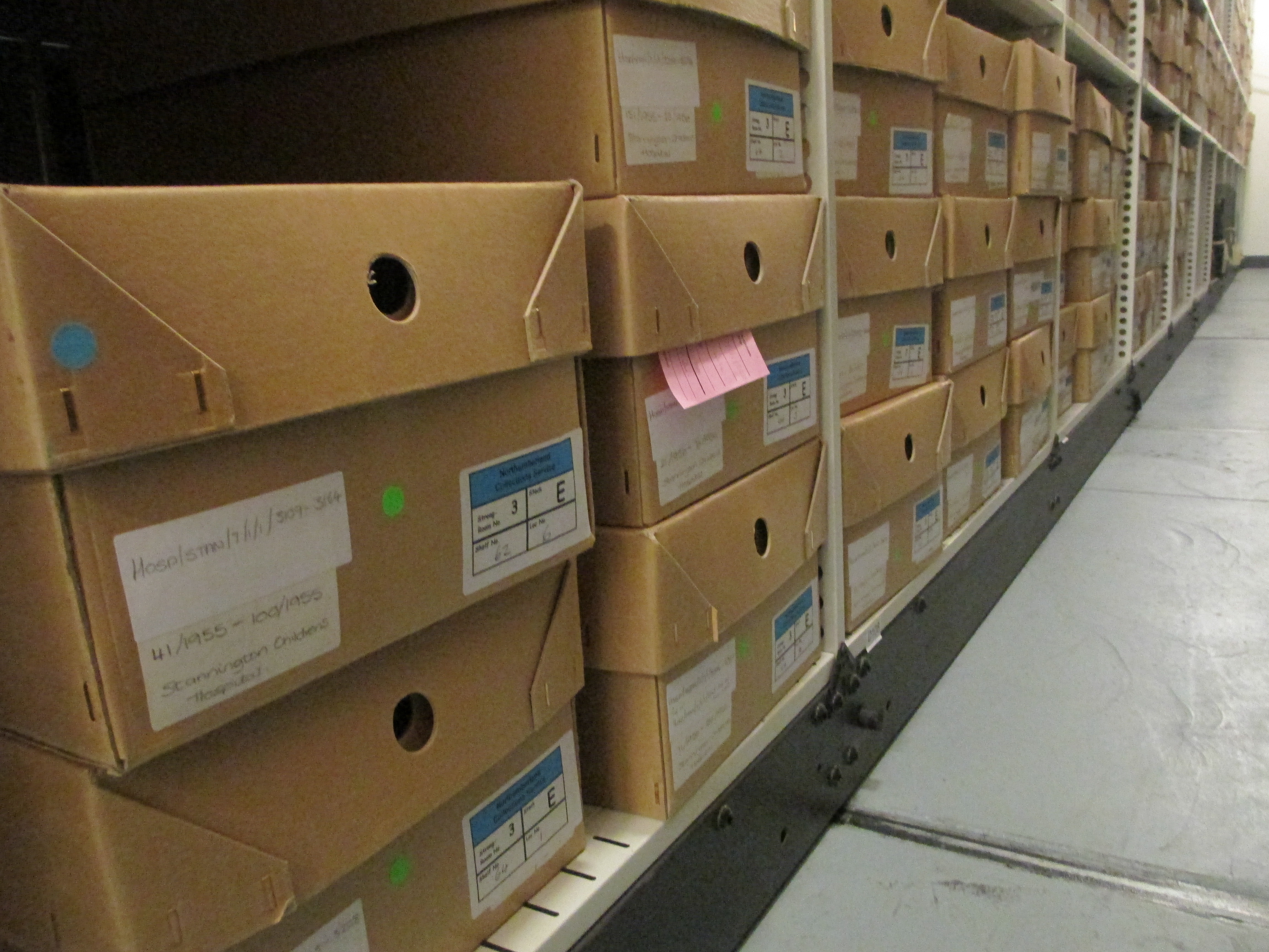
Over the course of the two phases of our Wellcome Trust funded project we have catalogued, conserved and digitised over 5000 patient medical files, as well as other records and photographs. In this time we hope we have taken an inaccessible, uncatalogued collection and made it accessible to study. In our final blog we will take a look back at what we have done in that time.

Some of the boxes of files waiting to be conserved early last year

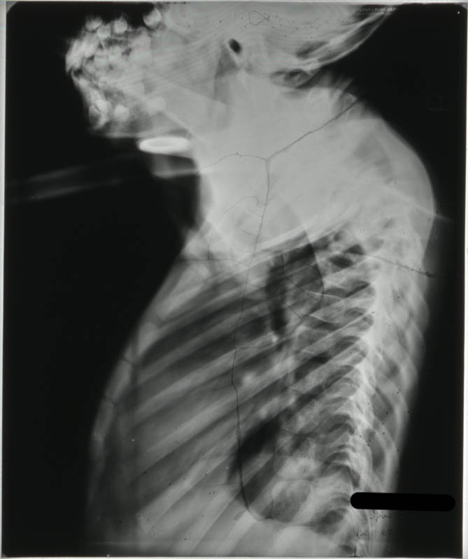
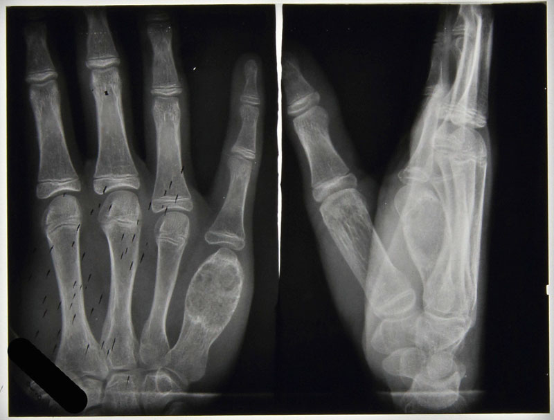
Phase one conserved and digitised 949 discharge books, catalogued these, the patient files and the rest of the collection, and digitised a whopping 14671 radiographs (taken of only 2245 of the patients!). The team also created our online exhibition, our exhibition banners, and attended conferences to publicise the collection. You can read more about the first phase in the team’s final blog here.

The radiographs being digitised

After the Wellcome trust kindly granted more funding for a second phase new team focused on the 4095 patient files, which had been catalogued.

A patient file conserved in its new acid-free cover
A patient file ready to be digitised

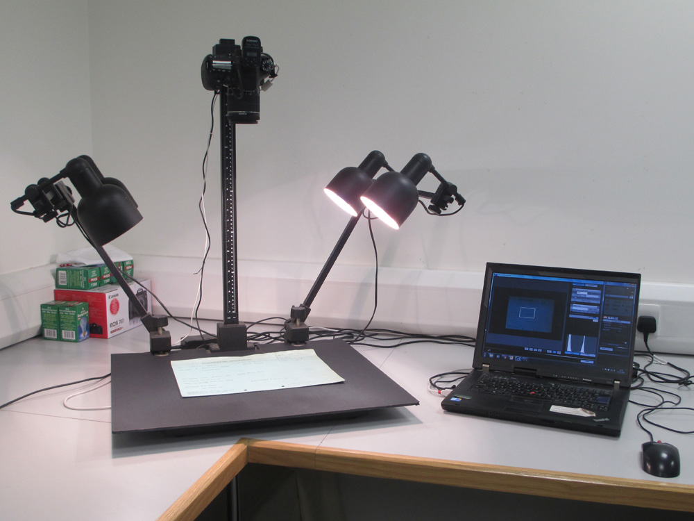
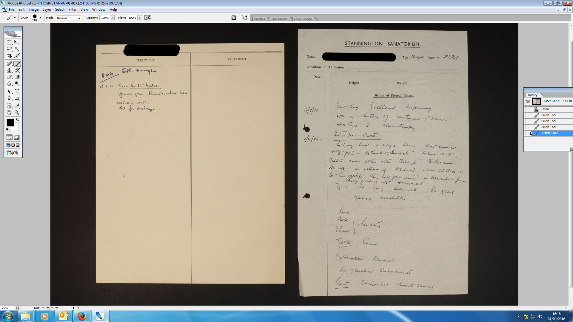
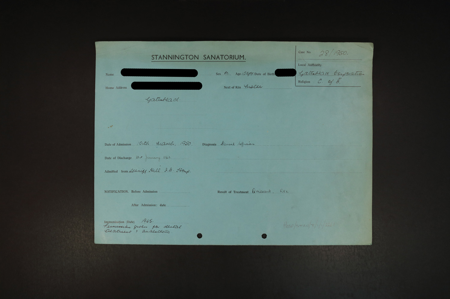
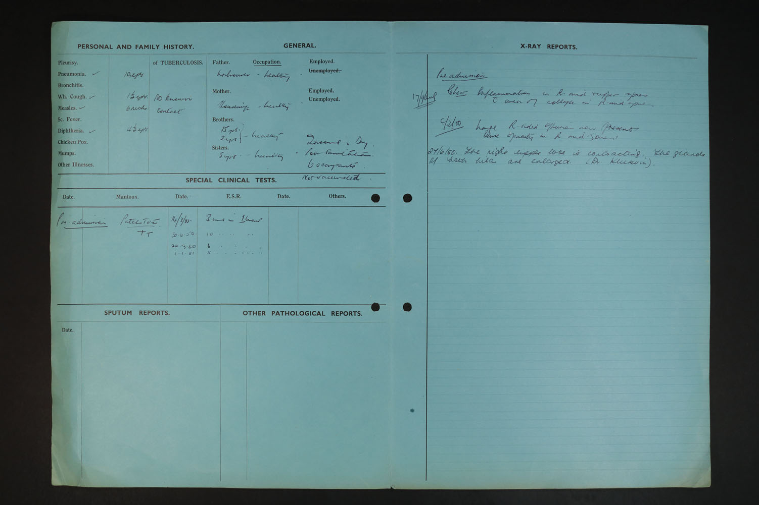
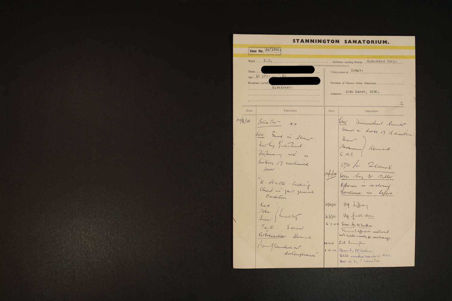
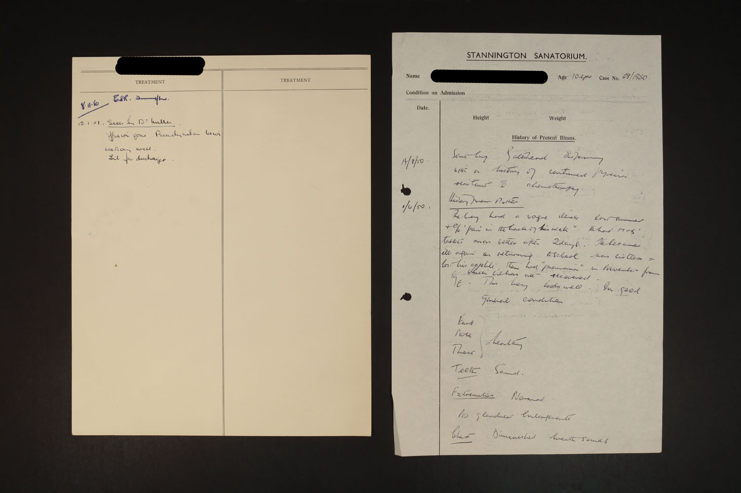
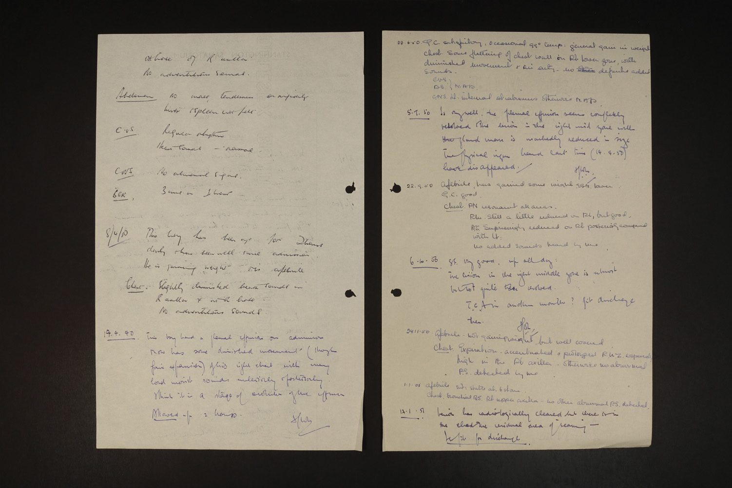
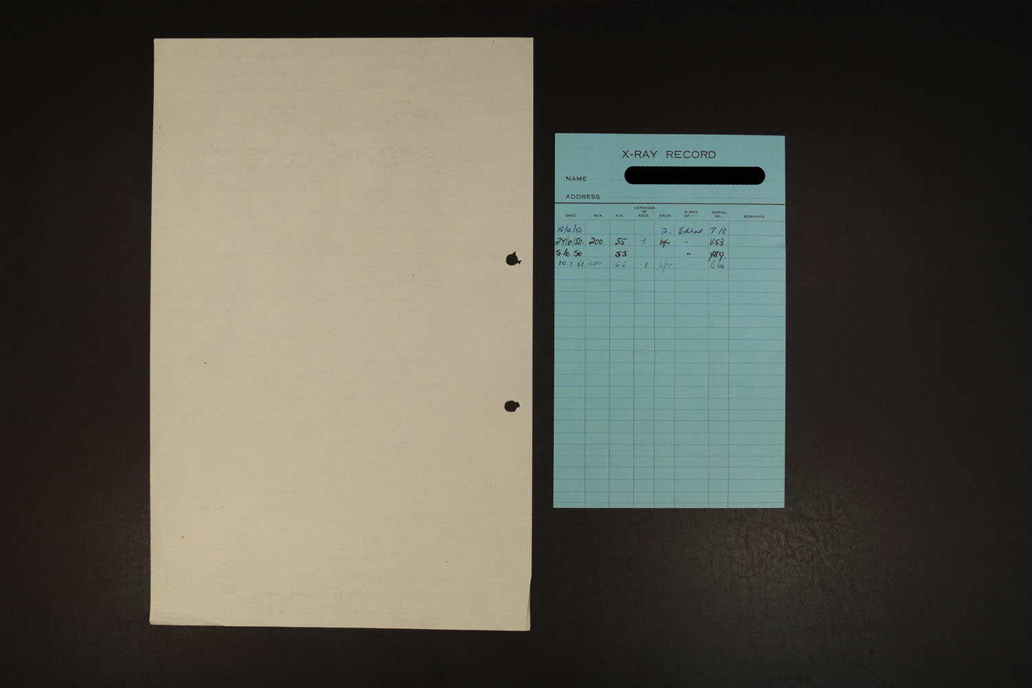
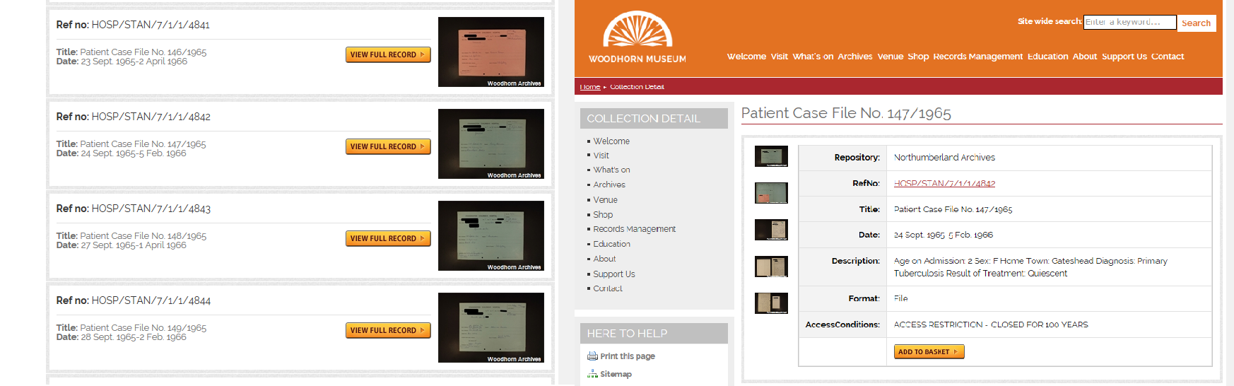
We conserved and digitised these, and redacted images were attached to our searchable online database. As they are closed records the names and addresses cannot be released, but this enables those researching the disease to access medical information about the cases for the ‘core’ documents. Here is how these look.

Print screens of the search and reference detail

As we could not digitise the entire files in each case the team decided to upload some examples on Flickr. Though there is no such thing as a ‘typical’ or ‘average’ Stannington file, we tried to choose a few redacted examples of the patient files that gave an impression of the collection as a whole, and the type of documents found within them. You can view these here.

The work began in phase one to raise awareness of the collection has continued. We have continued to blog about our progress and discoveries we have made about the collection, the sanatorium itself, and new collections that relate to it, such as May Brown’s the Atkin family’s photographs. There has been academic interest in research projects using the collection from a number of universities, which we hope to will continue to grow.

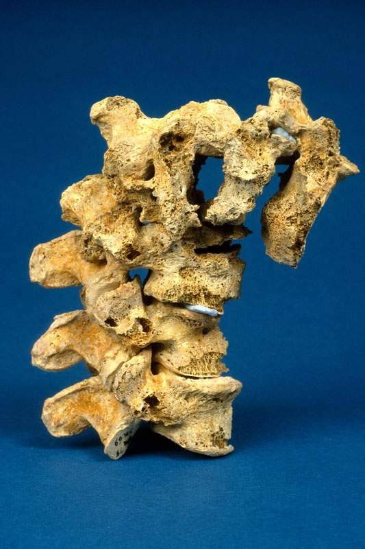
Our former colleague Rebecca Cessford is using the collection of radiographs and patient files in her PhD thesis to look at the potential as an aid to diagnose the disease in skeletal remains. You can find out more about her research in the blog she contributed to us on World Tuberculosis Day.

Margaret Wilkinson (right) with Dame Sian Philips at the recording of the play.

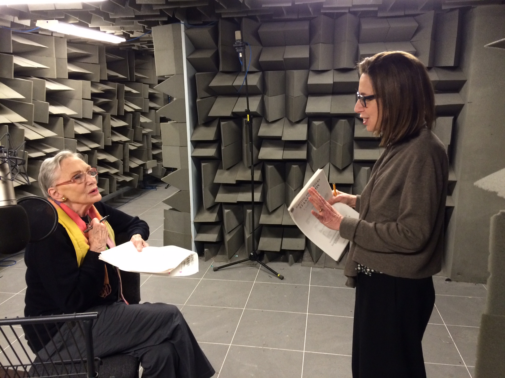
We also worked with playwright Margaret Wilkinson, who used the collection and our research to write a radio play for BBC Four’s Writing the Century. The play was based on the experiences of former Stannington Sanatorium nurse Marjorie Wilson. Margaret established the scene with Marjorie’s reminiscences, and used excerpts from letters within the patient files. The play was a great success, and listeners found the performance very moving. You can read more about Margaret’s inspiration in writing the play in the blog she wrote for us, and you can listen to the play through Audioboom.

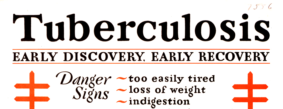
We hope to continue further interest in the collection by launching an education resource, now available through our website. In this we have laid out a little summary of the disease, history, treatment and the records at Stannington, and what life was like for staff and patients. This can be used as a resource for teachers to dip into in developing lessons, but also for older students to use for themselves.

The exhibition on display at St. Mary’s Church, Stannington.

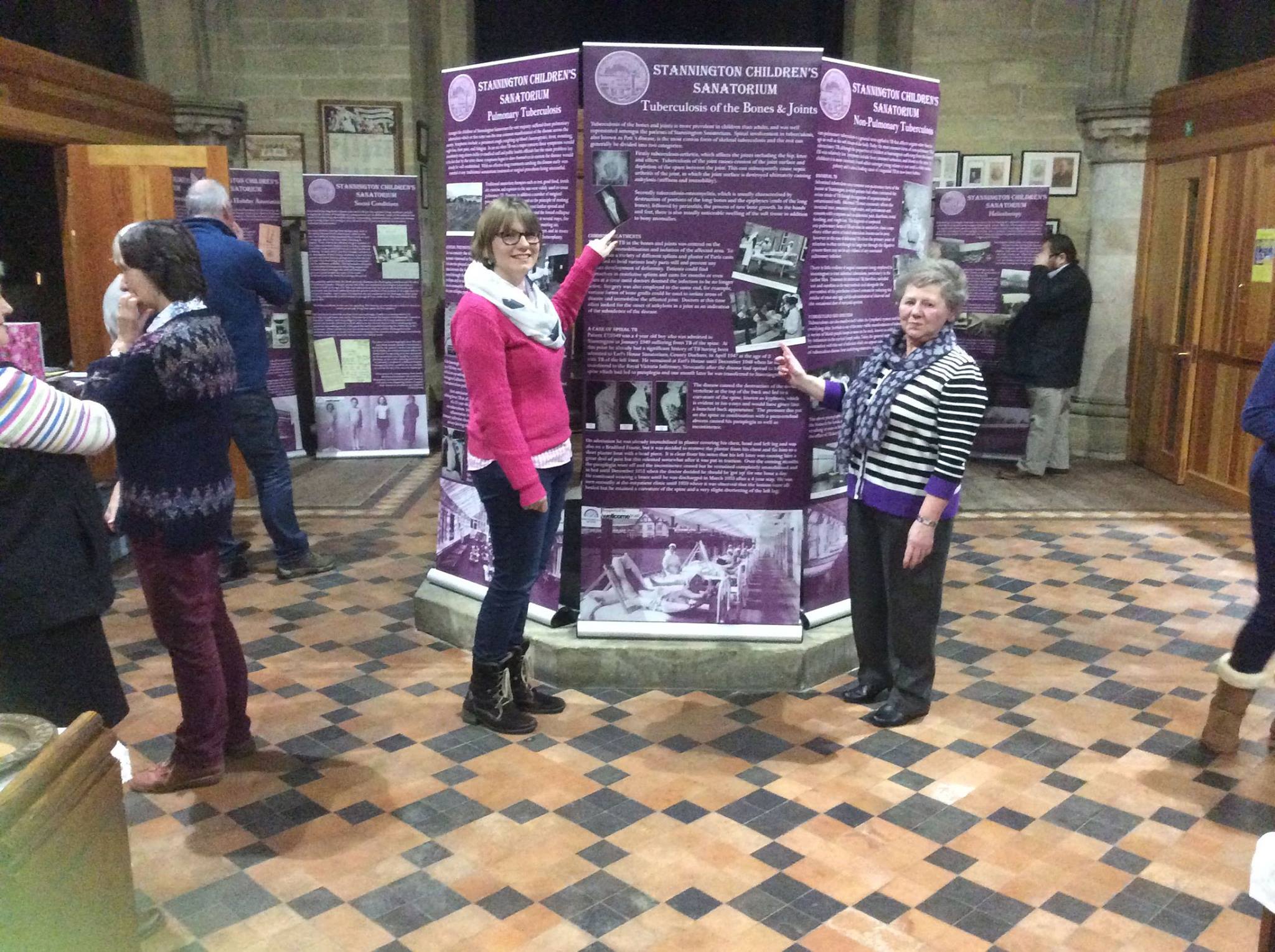
Recently the team have also set up our collection of banners at St Mary’s church in Stannington, with the Stannington History Group. It was a wonderful finish to the project to see them displayed so close to the sanatorium itself, with locals and visitors finding out more about the story of Stannington Sanatorium. We hope to tour the exhibition more throughout this year.

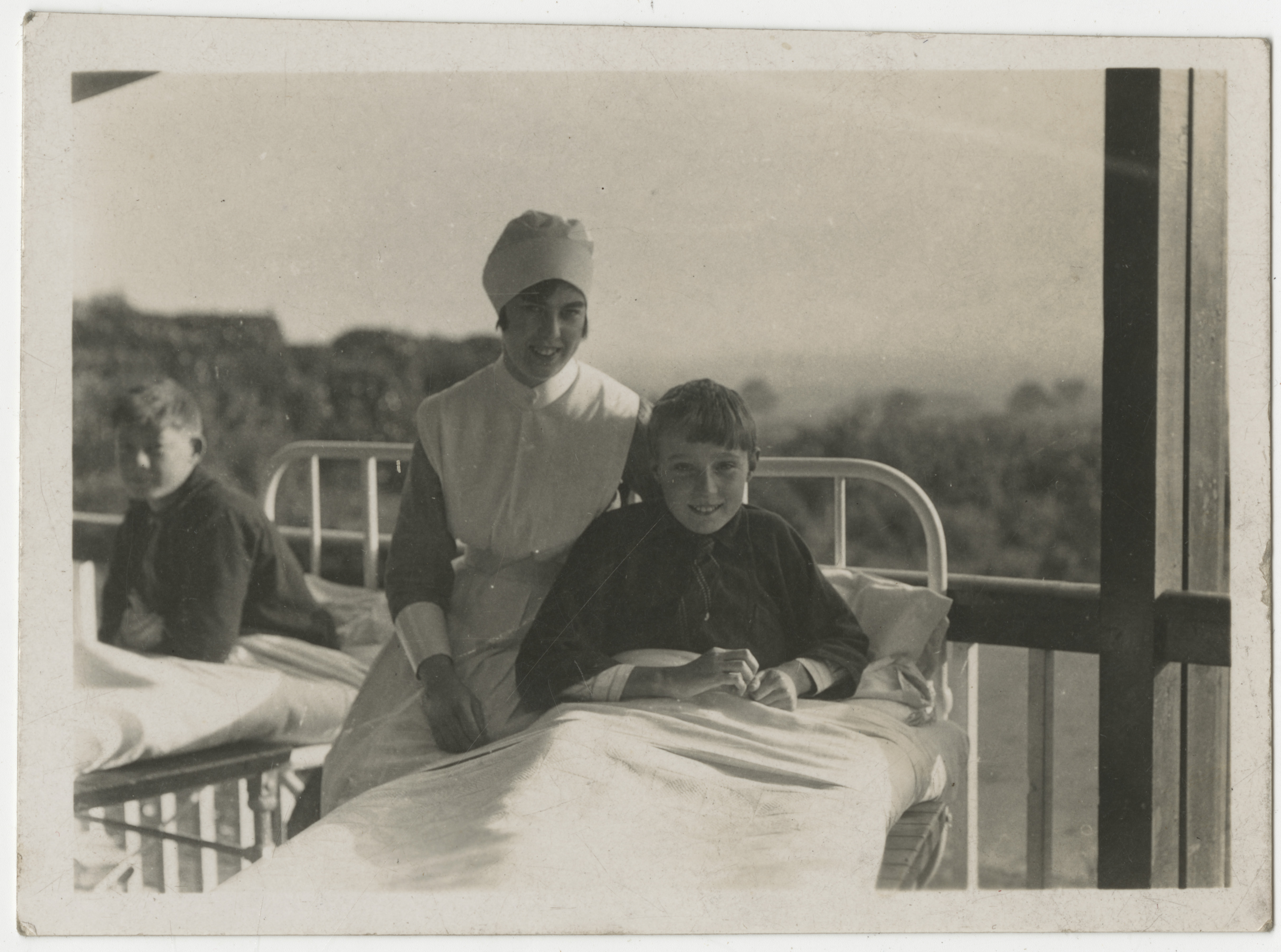
The project has revealed the rich value of the collection in terms of its research potential, as it contains files with detailed medical information and an excellent radiographic record. However it is also rich in the stories of the staff and patients who were associated with the sanatorium, many of whom have recounted their experiences to us during its course. We feel proud to continue to care for the documents, and feel it is important to continue to raise awareness. Far from this being the end of our research with the collection, this is a beginning of a new and more accessible way of learning from them.

The collection itself is housed at Northumberland Archives, and though a hundred year closure period is still in place (from the date of the document’s creation), former patients and some immediate family can still seek permission to access the patient files. If you are interested in accessing the collection for research purposes (we have statistics on the records as well as the files themselves), are interested in loaning our banners exhibition, or have any other queries about the collection please don’t hesitate to contact us at archives@northumberland.gov.uk

NRO 11036/3, one of May Brown’s photographs

If you are interested to know more please have a look at:

Our online exhibition

Our Flickr sets

World Tuberculosis Day 2016!

In honour of World Tuberculosis Day, we have a guest blog from Rebecca Cessford. Rebecca is a PhD researcher with the AHRC funded Heritage Consortium based at the Universities of Hull and Bradford. She will be using the Stannington Sanatorium Collection to study tuberculosis in the past using the archaeology of human remains and medical history. Here she tells us about her research and the role of the Stannington Collection in it.


When we think of tuberculosis (TB), images are conjured of a romantic disease causing a bloody cough, a pale complexion and weight-loss, the romanticised disease of the 19th century. What we do not think of is TB roaming the streets today. But tuberculosis is still a great threat, with over a million people dying of the disease each year and over 6,500 new cases declared in the UK during 2014. With increasing multidrug resistant strains of tuberculosis, is it possible to look back at a time before antibiotic drug therapy to better understand the future of this global emergency?

Early Discovery, Early Recovery 1929. Image from the National Library of Medicine, USA


Tuberculosis is a disease that extends as far back as the Neolithic period in Europe, with the earliest case reported in England coming from Dorset dating to the Iron Age. However, our ability to identify tuberculosis in skeletal remains from archaeological contexts is difficult. Firstly, tuberculosis of the bones and joints only affects 3-5% of all cases. Secondly, bone can only react to disease in a limited number of ways with many diseases causing similar bony destruction and remodelling. There are also problems identifying tuberculosis in the remains of children, due mainly to the under-representation of children in the archaeological record.

The most characteristic feature of tuberculosis in the skeleton is Pott’s Spine, an angular deformity in the mid to lower spinal column caused by the collapse of one or more vertebral bodies. The presence of this deformity has, for many years, been the only way of diagnosing tuberculosis in human remains with any certainty, despite the fact that any bone in the body can be affected. Advances in ancient DNA and biomolecular studies in archaeology mean tuberculosis can be tested for, even in the absence of any physical pathologies. However, these destructive and costly procedures are not without their limitations, still leaving much reliance on routine macroscopic observations (seen with the naked eye) of dry bone remains.

Pott's Spine the main diagnostic feature of tuberculosis in skeletal remains. Image courtesy of https://www.dur.ac.uk/images/archaeology/researchprojects/Roberts_TB.jpg
Pott’s Spine – characteristic collapse of the vertebral bodies causing an angular deformity of the spine due to tuberculosis. Image from University of Durham.


My research aims to look at the potential for using pre-antibiotic clinical radiographs (x- rays) as an aid to the macroscopic identification of tuberculosis in human remains, focussing specifically on infants and children. To do this, I intend to undertake a thorough examination of all the radiographs demonstrating skeletal tuberculosis to look at variations in progression of disease over time; the outcomes of healing on bones and the distribution of tuberculosis across the body where more than one bone was involved. In addition to this I will look at the corresponding medical file for each set of radiographs drawing on details outlined in the medical notes and x-ray reports to add to my own observations from the radiographs for an informed review of the underlying processes to bone and soft tissue being observed. It is hoped that the compilation of this data will provide a more detailed understanding of the processes involved in advancing tuberculous infection with comparative examples from pre-antibiotic radiographs. This strives to increase the ability to diagnose tuberculosis in archaeological remains even in the absence of Pott’s Spine.


Tuberculosis of the Knee: HOSP-STAN-07-01-02-91_09

Tuberculosis of the Spine - HOSP-STAN-07-01-02-1662-22
Tuberculosis of the Spine – HOSP-STAN-07-01-02-1662-22

Tuberculosis affecting the finger bones: HOSP-STAN-07-01-02-641_07


By studying the patterns of tuberculosis in the past we are better informed when it comes to dealing with the disease in the present and in the future. To be able to offer an evidence-based and informed approach to tackling tuberculosis we need better criteria for diagnosing it macroscopically in archaeological human remains, to get a more encompassing view of the various manifestations associated with it. The outcomes of my research will aim to act as an aid to the identification and study of tuberculosis in children in relation to archaeological remains further identifying the worth of pre-antibiotic medical records.

The Stannington Collection is a unique resource for studying this long standing infectious disease in children from the early to mid-20th century, many of which are still alive today living with the memories and/or side effects of the disease. I would also like to take this opportunity to thank the former patients of Stannington Sanatorium who expressed support for academic research to be undertaken on the collection during the first phase of the Stannington Sanatorium Project; their support makes research all the more worthwhile.

Digitising the Stannington Sanatorium patient files

As the Stannington Sanatorium Digitisation Assistant I am responsible for digitising over 4,000 patient case files and their contents, redacting personal identifiable information from these and uploading digital copies of the core documents to our publicly accessible, searchable, online catalogue. Before I can do this, my colleague, the Project Assistant has already sorted the forms, charts and various other items contained in each file into core and non-core documents and repackaged them in acid free folders for long term preservation. You can read more about this here.

A file ready to be digitised
The documents of patient file HOSP-STAN 07/01/01/2261 prior to digitisation

Before being digitised the pages in each file are checked to make sure they are in the correct order and any dog eared corners or folds are straightened out. The pages in the folder are then photographed on both the front and reverse sides to make sure that all the information in each folder is captured. The number of images captured varies from file to file and can be from just a few up to around fifty. Most files have around 10 to 15 pages, but the largest digitised to date contains over 140 pages!

We digitise the files using a high specification digital SLR camera connected to a computer. This allows us to take high resolution photographs of each page and is much quicker than digitising each page separately on a flatbed scanner. Using a flatbed would be the normal approach however the time constraints on the project mean we are digitising over 1,500 pages every week so this approach isn’t feasible.

Camera stand and laptop
The camera stand and laptop used to digitise each file.

The images are then processed and saved to nationally recognised standards set by The National Archives. Each file is saved in Tiff and Jpeg formats. The Tiff format creates files of a large size, but ensures that all the original information from the photograph is retained. Because of this and the sensitive nature of information in the un-redacted files they are stored on DVD in the Northumberland Archives strong rooms for security and long term preservation. The smaller Jpeg format allows us to keep these digital files on our own servers and readily accessible to staff and researchers if required.
The core documents from each folder (cover, case notes, x-ray card and discharge report) then have any information which could identify patients or is considered sensitive removed. This process involves using photo editing software on a computer, and reading each page to check for information such as names, addresses and dates of birth, and redacting it. This is a time consuming process not made easier by having to read a lot of hand written doctors’ notes!

Core documents being redacted in Adobe Photoshop
Adobe Photoshop is used to redact sensitive information from the core documents.

The final stage of the process is to upload the images to the relevant records on our online catalogue. Once this has been done a selection of pages from each file are publicly viewable along with each file’s catalogue entry and the digitisation process is complete. Currently over 18,000 pages have been digitised from over 1,400 patient files. Over 6,000 core documents are already viewable online via our catalogue in addition to a large number of radiographs and early patient files which were digitised during the first phase of the project.
A typical set of core documents from the file of a patient suffering from tuberculosis admitted to Stannington Sanatorium in the early 1950s can be seen below.

The front cover of a file
HOSP-STAN 07/01/01/2261/01

Inside the cover of a patient file
HOSP-STAN 07/01/01/2261/02

HOSP-STAN 07/01/01/2261/07 -
HOSP-STAN 07/01/01/2261/03

HOSP-STAN 07/01/01/2261/04

HOSP-STAN 07/01/01/2261/05

HOSP-STAN 07/01/01/2261/06

Pages from a patient file incuding discharge report
HOSP-STAN 07/01/01/2261/07