We touched upon the problems of infected milk supplies in a previous posting on abdominal TB and we’ll focus on the issue in more detail here. Mycobacterium bovis is the pathogen responsible for the development of TB in cattle, which is commonly referred to as bovine TB. The consumption of milk from cows infected with bovine TB and in turn the ingestion of mycobacterium bovis can lead to an individual developing TB. This was for many years a very common cause of TB in humans and remains so in countries that do not routinely pasteurise milk. Pasteurisation involves the heating of the milk in such a way as to kill off any bacteria that might be present, and through its use the spread of bovine TB to humans has nearly been eradicated in the UK.
The 1875 Public Health Act made it compulsory for local authorities to appoint a Medical Officer of Health (MoH) who produced an annual report detailing any health and sanitary issues in the district as well as giving a wealth of statistical information related to birth and death rates, population, infectious diseases and causes of death. The MoH for Northumberland makes regular reports on the situation in the county regarding tuberculosis including comments on the causes of abdominal tuberculosis and efforts made to prevent its spread. In the 1906 report he states:
“That about 30 per cent of the milch cows in England are tuberculous, and that consequently infants and persons suffering dangerous illness are in many cases being fed milk containing the organisms of tuberculosis” [NRO 3897/3, 1906 p.21]
The problem of infected animal products is clearly recognised by medical and sanitary officials early on in the 20th century but little is done to tackle the situation head on and so abdominal tuberculosis continues to be a significant problem. Three years later in 1909 the MoH expresses his frustration at the situation and lack of power to change it:
“The elimination of tuberculosis from dairy herds is a matter of great difficulty since, at present, no assistance is given, by the state, to the farmer who, for the benefit of the general public as well as for his own advantage, may wish to obtain a herd free from this disease.” [NRO 3897/3, 1909 p.33]
It is not until the 1940s that significant steps were taken to introduce tuberculin tested milk and encourage pasteurization.
“The eradication of tuberculosis from our milk supplies is a matter of greatest importance to us all, and it is encouraging to note the marked increase in the production of milk from tuberculin tested cows. 45% of all the milk produced in the County was from such herds, and it is known that in 1948 the proportion had risen to more than 50%.” [NRO 4081/1, 1947 p.8]
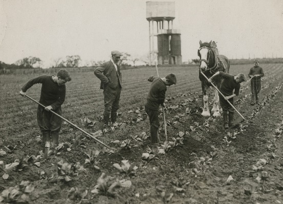
Boys at work on the farm
Milk supplies were something given great consideration by those responsible for the establishment of Stannington Sanatorium from the outset. In 1905, two years before the official opening of the sanatorium, a farm colony was established on the site to take in young boys and provide them with training. It was from here that the sanatorium was able to receive a safe supply of milk from tuberculin tested cows. Tuberculin testing is another method used in preventing the spread of bovine TB whereby the cows were tested to see whether they carried mycobacterium bovis rather than treating the milk itself. This method was used quite commonly early on before the onset of widespread pasteurisation and would have been essential to the recovery of many of the patients and in preventing any of them acquiring any further infection. As time goes on, and tuberculin testing and pasteurisation is implemented more widely across the county, it is notable when looking at the patient files that instances of abdominal TB decrease particularly as we enter the 1950s.
Sources:
ALLISON, T. M. (1908) Children’s Sanatorium, Stannington, Northumberland, British Journal of Tuberculosis, 2 (3), p.204
SCHOFIELD, P. F. (1985) Abdominal Tuberculosis, Gut, 26 (12), pp.1275-1278
NORTHUMBERLAND ARCHIVES: NRO 03897, Northumberland County Council: County Medical Officer of Health Reports, 1893-1935
NORTHUMBERLAND ARCHIVES: NRO 04081, Northumberland Health Authority: Records, 1942-1970