World Tuberculosis Day 2016!

In honour of World Tuberculosis Day, we have a guest blog from Rebecca Cessford. Rebecca is a PhD researcher with the AHRC funded Heritage Consortium based at the Universities of Hull and Bradford. She will be using the Stannington Sanatorium Collection to study tuberculosis in the past using the archaeology of human remains and medical history. Here she tells us about her research and the role of the Stannington Collection in it.


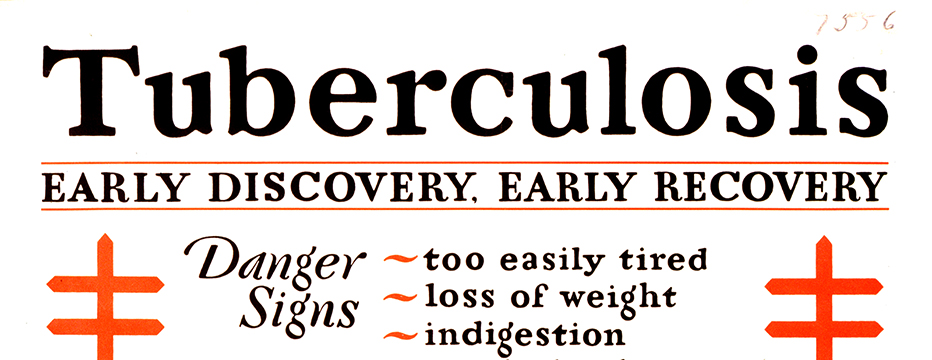
When we think of tuberculosis (TB), images are conjured of a romantic disease causing a bloody cough, a pale complexion and weight-loss, the romanticised disease of the 19th century. What we do not think of is TB roaming the streets today. But tuberculosis is still a great threat, with over a million people dying of the disease each year and over 6,500 new cases declared in the UK during 2014. With increasing multidrug resistant strains of tuberculosis, is it possible to look back at a time before antibiotic drug therapy to better understand the future of this global emergency?

Early Discovery, Early Recovery 1929. Image from the National Library of Medicine, USA


Tuberculosis is a disease that extends as far back as the Neolithic period in Europe, with the earliest case reported in England coming from Dorset dating to the Iron Age. However, our ability to identify tuberculosis in skeletal remains from archaeological contexts is difficult. Firstly, tuberculosis of the bones and joints only affects 3-5% of all cases. Secondly, bone can only react to disease in a limited number of ways with many diseases causing similar bony destruction and remodelling. There are also problems identifying tuberculosis in the remains of children, due mainly to the under-representation of children in the archaeological record.

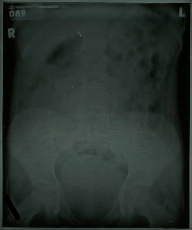
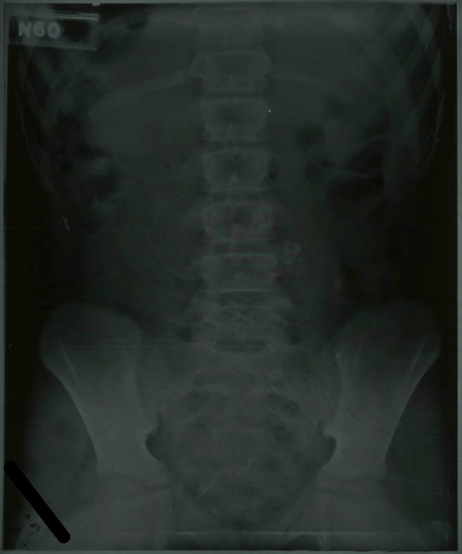
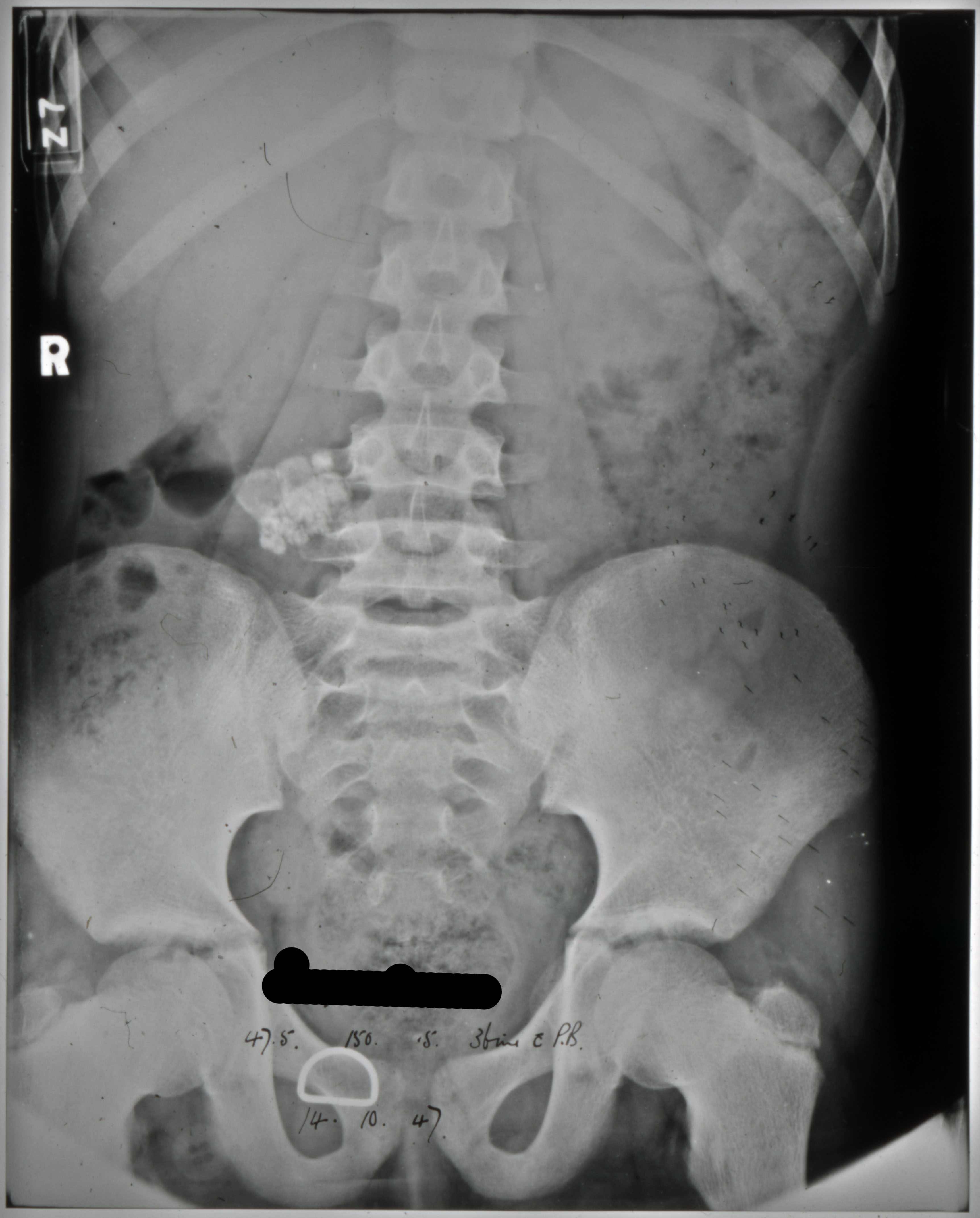
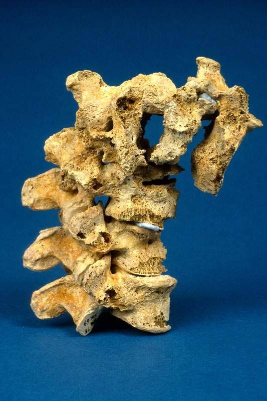
The most characteristic feature of tuberculosis in the skeleton is Pott’s Spine, an angular deformity in the mid to lower spinal column caused by the collapse of one or more vertebral bodies. The presence of this deformity has, for many years, been the only way of diagnosing tuberculosis in human remains with any certainty, despite the fact that any bone in the body can be affected. Advances in ancient DNA and biomolecular studies in archaeology mean tuberculosis can be tested for, even in the absence of any physical pathologies. However, these destructive and costly procedures are not without their limitations, still leaving much reliance on routine macroscopic observations (seen with the naked eye) of dry bone remains.

Pott's Spine the main diagnostic feature of tuberculosis in skeletal remains. Image courtesy of https://www.dur.ac.uk/images/archaeology/researchprojects/Roberts_TB.jpg
Pott’s Spine – characteristic collapse of the vertebral bodies causing an angular deformity of the spine due to tuberculosis. Image from University of Durham.


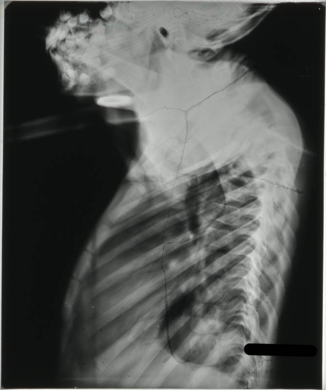
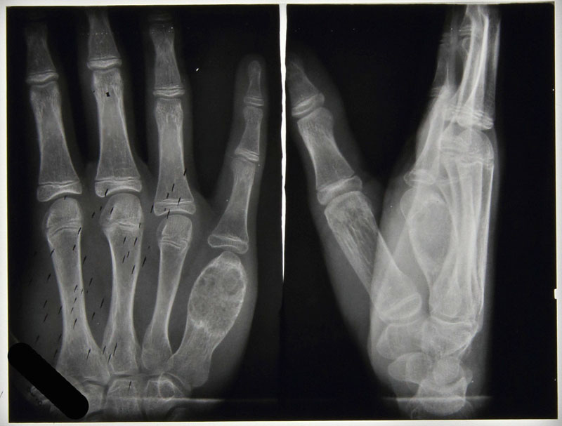
My research aims to look at the potential for using pre-antibiotic clinical radiographs (x- rays) as an aid to the macroscopic identification of tuberculosis in human remains, focussing specifically on infants and children. To do this, I intend to undertake a thorough examination of all the radiographs demonstrating skeletal tuberculosis to look at variations in progression of disease over time; the outcomes of healing on bones and the distribution of tuberculosis across the body where more than one bone was involved. In addition to this I will look at the corresponding medical file for each set of radiographs drawing on details outlined in the medical notes and x-ray reports to add to my own observations from the radiographs for an informed review of the underlying processes to bone and soft tissue being observed. It is hoped that the compilation of this data will provide a more detailed understanding of the processes involved in advancing tuberculous infection with comparative examples from pre-antibiotic radiographs. This strives to increase the ability to diagnose tuberculosis in archaeological remains even in the absence of Pott’s Spine.


Tuberculosis of the Knee: HOSP-STAN-07-01-02-91_09
Tuberculosis of the Spine - HOSP-STAN-07-01-02-1662-22
Tuberculosis of the Spine – HOSP-STAN-07-01-02-1662-22
Tuberculosis affecting the finger bones: HOSP-STAN-07-01-02-641_07


By studying the patterns of tuberculosis in the past we are better informed when it comes to dealing with the disease in the present and in the future. To be able to offer an evidence-based and informed approach to tackling tuberculosis we need better criteria for diagnosing it macroscopically in archaeological human remains, to get a more encompassing view of the various manifestations associated with it. The outcomes of my research will aim to act as an aid to the identification and study of tuberculosis in children in relation to archaeological remains further identifying the worth of pre-antibiotic medical records.

The Stannington Collection is a unique resource for studying this long standing infectious disease in children from the early to mid-20th century, many of which are still alive today living with the memories and/or side effects of the disease. I would also like to take this opportunity to thank the former patients of Stannington Sanatorium who expressed support for academic research to be undertaken on the collection during the first phase of the Stannington Sanatorium Project; their support makes research all the more worthwhile.

The Stannington Radiographs

The radiographs make up a significant part of the Stannington collection with a total of 14,674 separate images relating to 2220 different patients covering roughly a 20 year period from 1936 to c.1955. When the records were recovered in the 1980s the vast majority of the radiographs were copied on to microfiche and the originals destroyed as they were unstable. However, we still have 326 original radiographs within the collection. Over the course of the project all the microfiche images and the originals will be digitised and made publicly accessible. We also hope to preserve the remaining original radiographs as examples of how x-ray images at the time were produced. The problem here lies with the unstable nature of the film and its natural degradation.


All the radiographs were produced on cellulose acetate film, known as safety film as it replaced the earlier nitrate film that was highly flammable and potentially self-combustible, a problem for many film archives today. Over time the cellulose acetate film naturally breaks down, the early stages of this are recognisable by the strong smell of vinegar coming off the film as the process gives off acetic acid and because of this is known as vinegar syndrome. Eventually as the base of the film and the top layer pull away from one another the film will begin to buckle and crack and bubbles can form under the surface.

IMG_0904
Figure 1
IMG_0905
Figure 2

This process is already evident in several of the radiographs we hold (figures 1 & 2) and unfortunately there isn’t anything that can be done to reverse or halt the process. By storing the films in a closely monitored temperature and humidity controlled environment we hope to delay the process in most of the radiographs for as long as possible.


IMG_0903
Figure 4
IMG_0902
Figure 3


The x-rays were originally stored in ordinary brown envelopes and there could be as many as 15 in each envelope. (Figure 3)  To help preserve them we have instead transferred each x-ray into an individual acid free sleeve. (Figure 4)  By storing them individually we are able to minimise any accumulation of acetic acid that is produced in the degradation process.  Thanks to advice from conservators at Durham County Record Office and their assistance in sourcing the new x-ray envelopes, all the original films are now safely stored in their own envelopes in our photogrpahic strong room.


This degradation is evident in some of the microfiche as well as the original films had obviously started to buckle already at the time they were transferred to microfiche in the 1980s. Consequently some of the images are obscured by a crackling effect. Nevertheless the vast majority of the 14,674 images remain easily readable and the digitisation process will mean that they remain clearly accessible for future use.


[See our Flickr stream for examples of some of the radiographs https://www.flickr.com/photos/99322319@N07/sets/72157648833066476/]

Case Study – Abdominal Tuberculosis

Abdominal tuberculosis was a common diagnosis amongst the patients of Stannington Sanatorium and the patient case notes and radiographs give some indication as to the progression and manifestation of this form of TB.  As was seen in the post from 19 November detailing the different types of TB, abdominal tuberculosis was a common extra-pulmonary form of the disease in which patients had often contracted the bovine strain of tuberculosis (mycobacterium-bovis) through the ingestion of unpasteurised or contaminated milk.  We will explore the problems that arose from contaminated milk in a later post.

Abdominal TB most commonly affects the intestinal tract, mesenteric lymph nodes, and peritoneum and presents with symptoms such as abdominal pain, diarrhoea, rectal bleeding, and weight loss.  The diagnosis of suspected extra-pulmonary forms of TB can often be assisted by chest x-rays where either active or healed tuberculous lesions can be seen.  However in the case of abdominal TB where the primary point of infection is often not through the lungs but through the digestive system there may not be any evidence of any associated pulmonary infection. (Lambrianides et al, p.888)  We don’t see as many radiographs within the Stannington records relating to abdominal tuberculosis in comparison to other manifestations of the disease such as pulmonary or bones and joints but there are nevertheless some good examples throughout the collection.


Case Study 1

Figure 1 - HOSP/STAN/7/1/2/21 (2)
Figure 1 – HOSP/STAN/7/1/2/21 (2)
Figure 2 - HOSP/STAN/7/1/2/21 (3)
Figure 2 – HOSP/STAN/7/1/2/21 (3)


The above radiographs are some of the earliest examples of abdominal TB from amongst the Stannington collection.  They both relate to patient 80/21, a 9-year old female admitted in 1939 who presented with  “vague abdominal signs and pain and traces of albumin in urine,” and a report on an x-ray taken prior to admission states that “calcified glands visible in abdomen above sacrum.”  The reports given on the above two x-rays in the patient’s medical notes are very brief, with the first, figure one, being taken on 20 April 1939 and the report simply stating “gland to right of spine.”  The second, figure two, was taken on 12 October 1939 and the report stated “calcification better seen in abdominal glands.”


Case Study 2

NRO-3000-HOSP-STAN-07-01-02-1463-06
Figure 3 – HOSP/STAN/7/1/2/1463 (6)

Figure 3 relates to a patient who was originally admitted in June 1940 as patient 86/46 and discharged in May 1941 only to be re-admitted some years later in September 1947 as patient 118/1947.  On his first admission he was 5 years old and it was reported that during the past year he had had several attacks of vomiting and abdominal pain but an initial examination found no resistance in the abdomen and no mass was felt.  No glands were seen in the x-rays of his abdomen taken at this time.  The above radiograph was taken in October of 1947 following his second admission and the radiographic images from this slightly later period tend to be much clearer and better defined than those from the early 1940s, such as in figures 1 & 2.   When he was admitted the second time he presented with “listlessness, poor appetite, vague abdominal pain & night sweats” but during his stay no evidence of any active disease was actually found, with the above x-ray showing calcified abdominal glands, presumably as a result of his previous, now quiescent, case of abdominal TB from 7 years earlier.  He was discharged less than three months later once his symptoms had settled down.


There is little evidence of surgical treatments being employed in Stannington to treat abdominal tuberculosis, particularly in the earlier files.  This is corroborated by early literature on abdominal TB in children where rest and sunshine are cited as the main methods of treatment alongside the prevention of the putrefaction of bowel contents by reducing the intake of meat and eggs and the administration of charcoal and the occasional dose of mercurial aperient. (Sundell, 1926)  Later studies however recommend the use of surgical treatments to deal with intestinal lesions in order to prevent healing by fibrosis which could lead to obstructions causing later problems. (Kapoor & Sharma, 1988)  This healing process is evident in the radiographs of patient 118/1947 showing the calcified glands, with the possibility of problems occurring later on in life being something to consider with many of the Stannington patients.


Sources

KAPOOR, V.K. & SHARMA, L.K. (1988) Abdominal Tuberculosis, British Journal of Surgery, 75 (1), pp.2-3

LAMBRIANIDES, A.L., ACKROYD, N. & SHOREY, B.A. (1980) Abdominal Tuberculosis, British Journal of Surgery, 67 (12), pp.887-889

SUNDELL, C.E. (1926) Abdominal Tuberculosis in Children, Postgraduate Medical Journal, 2 (14), pp.24-26